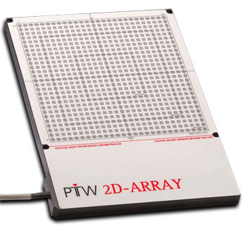
The 2D-ARRAY seven29 - Overview
Basic hardware description
The 2D-ARRAY seven29 is a two dimensional ionization chamber array with 729 vented ionization chambers, each having cubic dimensions. The small squares on the surface of the image show their position.
| Some technical parameters (from PTW manual) | |
|---|---|
| Measured values | Absorbed dose to water [Gy] |
| Measuring mode | Dose or dose rate |
| Maximum field size | 27 x 27 cm |
| Chamber voltage | 400 V |
| Measuring range (dose rate) | 500 mGy/min ... 10 Gy/min (resolution: 1 mGy/min) |
| Measuring range (dose) | 200 mGy ... 1000 Gy (resolution: 1 mGy) |
| Reproducibility | Better than +/- 0.5% (IEC 60731) |
| Linearity | Better than +/- 0.5% (IEC 60731) |
| Long term stability (ARRAY and interface) | Better than +/- 1% per year Better than +/- 1% after 1000 Gy |
| Dead time | N/A |
| Dimensions | ARRAY: 22 mm x 300 mm x 420 mm Interface: 80 mm x 250 mm x 300 mm |
| Weight | ARRAY: 3.2 kg, Interface: 2.4 kg |
| Recalibration | Recommended after 2 years |
| Detector type | Vented ionization chamber |
| Arrangement | Matrix of 27 x 27 chambers, 10 mm center to center |
| Chamber size | 5 mm x 5 mm x 5 mm |
| Reference point | 5 mm behind surface |
| Effective Point of Measurement | See our own measurements |
| Wall material | Graphite |
| Area density above the chamber volume | 0.6 g/cm2 |
Treatment room setup
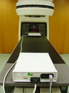
The array is connected to an interface by a 1.5 m cable. The interface is usually placed at the distal end of the treatment couch (Fig.1). From here, a RS232 cable (Fig.2) goes to the control room, where a common PC controls the measurements.
 |
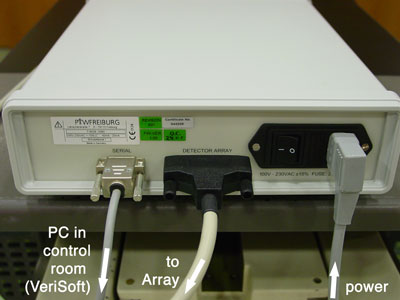 |
| Fig.1: ARRAY and Interface. | Fig.2: The back side of the Interface. |
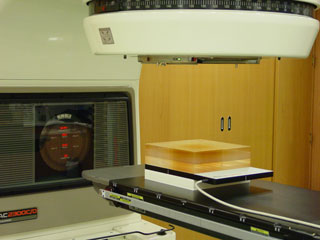
Fig.3 shows a typical setup, where a solid block of PMMA is used as buildup to simulate a depth of 10 cm in the patient. 4 cm of RW-3 are used as backscatter. This setup was CT-scanned once, the resulting 3D-phantom-patient is used in the TPS (Eclipse) as Verification phantom for IMRT fields (field-by-field verification).
 |
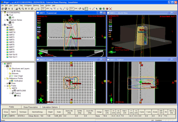 |
| Fig.3: Typical setup during IMRT verification. | Fig.4: Corresponding measurement. |
The ARRAY comes with a calibration certificate for 60Co. If the ARRAY is used for absolute dose measurements, the effective point of measurement P(eff) is, according to PTW, the entry window of the central chamber (#365), 5 mm below the surface (Fig.4). Own experiments have shown that it is about 1 mm deeper. This corresponds to a displacement correction of 1.5 mm from the center of the chamber in direction of focus. The cubic chamber corresponds more to a thimble chamber than to a plane parallel chamber (cubic size, no guard ring).
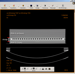
Fig.5 shows a scan of the ARRAY, embedded in Solid Water. Fig.6 shows the corresponding scout view.
 |
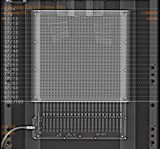 |
| Fig.5: CT-scan of 2D-ARRAY in Solid Water. | Fig.6: Topogram of the same setup. |
Basic software description
As soon as the array is set up on the treatment couch, the components connected an switched on, the measurements are controlled from outside the treatment room. We are using different PTW software, depending on the type of use. There is software available for basic measurements (MatrixScan), IMRT verification (VeriSoft) and constancy checks (MultiCheck).
The MatrixScan software per se is usually never started directly: if VeriSoft is used and one starts a 2D-ARRAY measurement, VeriSoft triggers MatrixScan to perform the measurement. If the result is accepted, the control goes back to VeriSoft. The same holds for MultiCheck. This operation is very smooth.
