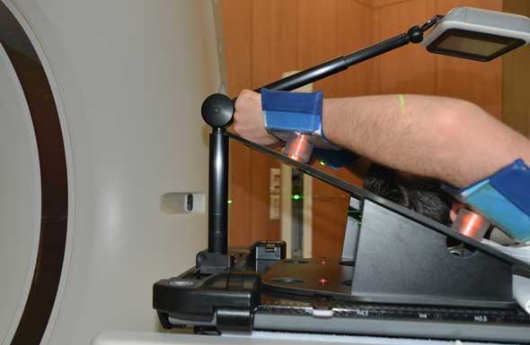
(Vienna, March 7, 2014): After removal of the RPM system on March 5, we installed Varian's revolutionary new Gating solution RGSC (Revolutionary Gating for SCanners1) on our Toshiba AquilionLB. Installation, testing and acceptance took 1.5 days, without any stress. On March 7, we tested the system clinically for the first time, using both integrated coaching possibilities: audio and video coaching.
Differences between RPM and RGSC
While the gating principle is still the same (reflective marker block on patient + camera), hard- and software are completely new.
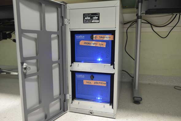
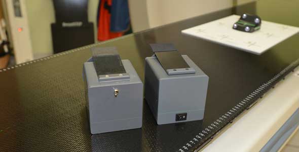
The major components of the well designed RGSC cabinet are two blue "shoe-boxes": the workstation (upper slot) and a real-time node (lower slot). The real-time node is always on, see the instruction labels on photo ("drehen" = turn, "abdrehen OK" = shut-down OK, "nix abdrehen" = do not turn off):
The cabinet also contains a 16 port switch, a Juniper firewall, and a power injector for a WLAN acces point. The latter is positioned in the CT room, has Power over Ethernet (PoE), and streams the visual coaching animation to the VCD (Visual Coaching Device), a cable-free LCD monitor. The VCD can be conveniently positioned in front of the patient's face:
This VCD comes with battery packs and a Varian charger. The VCD is on a telescopic arm, which is connected to the CT overlay with a couch mount that is specific for the carbon overlay.
The VCD can display various animations such as simple trail, a 1-D slider which should be kept within the gated range (the smiley is then displayed as "reward"), or a dog which has to be kept on its path (thereby collecting "targets" = bones):
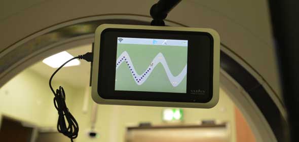
On the next photo, the VCD has a USB mouse attached. This is only used for diagnostics and to start the demo mode. Clinically, no cable is running to the VCD. The position of the reflector block is transferred via WLAN to the VCD, which then generates the animation (It's actually a little computer).
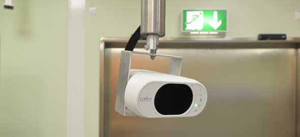
The Camera uses the same principle as the RPM camera, but is a complete redesign. A red guiding laser is hidden behind the front cover of the camera (similar to the laser on the TrueBeam NDI gating cameras). This laser can only be operated by removing the front cover. It should point to isocenter.
We have designed an aluminum frame and mounted the camera to the same roof mount as the RPM camera:
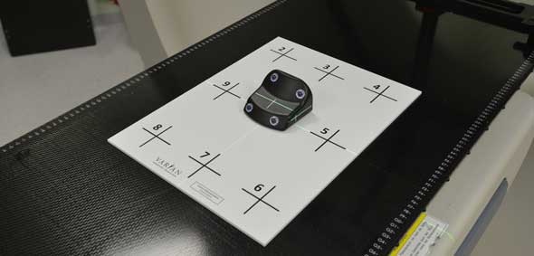
RGSC uses the same marker block (irregular shape, four reflective lenses) as the TrueBeam:
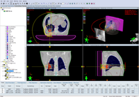
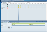
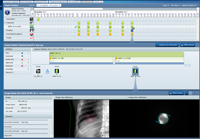
Tracking is always 3D. A special camera calibration plate facilitates recalibration, should it be necessary. Here are software screenshots for positions 1, 2, and after the last step. Reflections of the carbon couchtop can fool the software. This can simply be avoided, by placing a sheet of paper on the couchtop, in front of the block.
Calibration check is not mandatory any more. Recommeded check interval is 1 day.
Workstation software is visually designed like the TrueBeam software. Users log in via OSP. Several user rights can be set. Messages pop up. In this sense, RGSC is a "little TrueBeam".
Whereas RPM (last version was 1.7.5 Deluxe) was a stand-alone solution, RGSC (current version 1.0) is fully integrated into the ARIA system, if TrueBeam and ARIA 13 are used. But RGSC can also be used with C-Series Clinacs (where RPM is installed on the Clinac), or with TrueBeam and ARIA 10 or 11.
The only part which is still the same (except for the new on/off switch) is the breathing phantom provided by Varian. This device simply cannot be improved!
For real phantom scans, we use our Quasar respiratory platform. One simple phantom which can be placed on the platform is the IGRT cube from CIRS. The platform can be moved around using sinusses or real patient waveforms.
First Experiences
Although it is version 1.0, the whole RGSC system appears to be very mature and stable. No problem of any kind was encountered during installation and during the first day of clinical use.
Update, March 2015: Last month, Toshiba installed a new serial interface between RGSC and the Aquilion LB, which is currently under testing in our institute. The benefit: after a 4D scan (and before the retrospective reconstruction is started on the CT console), it is not necessary any more to compare the breathing patterns peak-by-peak on the gating system and the scanner. This greatly improved our workflow!
Notes
1Just joking. R stands for Respiratory, of course.
2For breast, a kV-kV image pair is usually only acquired during the first session. But because the MV-before-image showed a large shift, the kV-kV-image pair was also acquired in the second session.
