Preparing the Eclipse Water Phantoms for EPIQA Calculations
EPIQA assumes that the planar dose distributions are calculated in one of the following geometries:
- If Gantry and Couch are at zero angle, it is called the (0, 0) geometry.
- It is also possible to calculate at (90, 90), which means that Gantry and Couch are rotated.
In either case, for each geometry at least one water phantom first has to be created in Eclipse. We will use the (0, 0) geometry for our examples. The (90, 90) geometry only has some advantages if the PBC algorithm is used.
To make sure that always the correct phantom is selected during a verification session, we find it convenient to have the geometry written in the name of both the phantom and the structure set (red arrows):
What do these structure sets contain?
At KFJ, we have the old Varian R-arm, which means that we measure EPID dose at a Source Detector Distance (SDD) of 105 cm. Depending on setup (MIX or DMAX) and energy (6, 15MV), we need different phantom gometries. The comment in the above screenshot tells it all: the MIX structure set for 6MV provides a field SSD of 103.5 cm. At 1.5 cm depth (in other words, at 103.5 + 1.5 = 105 cm from focus), we had generated the MU/Gy tables of EPIQA (="calibrated EPIQA"). We did not measure MU/Gy with chamber, but calculated the doses in Eclipse.
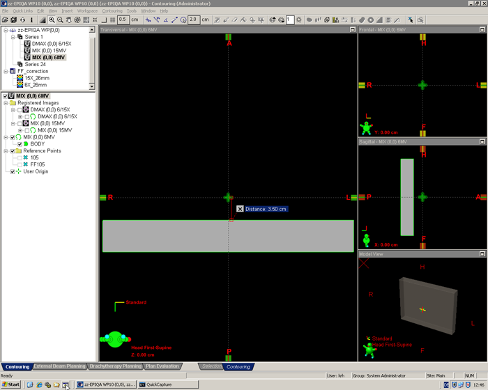
A lot of mouse clicks and keystrokes can be saved if the BODY of the phantoms is positioned cleverly: If the BODY structure is placed somewhat below the image center (Eclipse puts the User Origin there), all further verification plans which use this phantom will have the fields positioned in the correct distance automatically. The user only has to make sure that the dose planes are exported at 5 cm below isocenter (SDD 105 cm, Y = 5.00 cm).
This is the 6MV phantom for MIX mode:
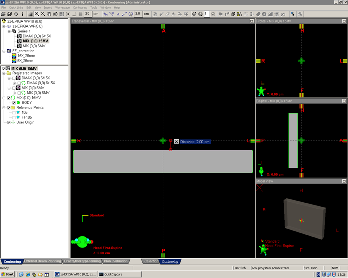
For 15MV MIX mode, we calibrated EPIQA at 3 cm depth. This is a little deeper than the real dmax (2.7 cm), but as long as the calibration geometry and the setup geometry are consistent, it is correct. The 15MV structure set for MIX mode has the BODY placed 2.0 cm below the User Origin, which means that the fields will be automatically placed at an SSD of 102 cm:
By the way: Our virtual EPID has a size of about 40 x 30 x 5 cm, which is large enough for all IMRT fields. The image slices have a thickness of 1 mm, the block is filled with water (0 HU).
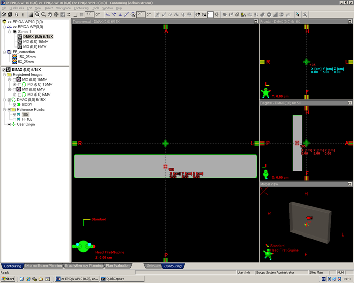
Our last phantom is for DMAX geometry, which means that additional buildup has to be placed on the EPID at the time of image acquisition. We have a 40 x 30 cm slab of PMMA for this, which is 15 mm thick. With a water equilvalent thickness of about 18 mm plus the 8 mm intrinsic water equivalent depth of the EPID, we get 2.6 cm of water. This is a little less than the real dmax for 15MV, but again: it is EPID calibration depth, and depths only have to be used consistently.
The same 15 mm slab of PMMA is used for 6MV and 15MV. Therefore, when using DMAX mode, dose planes in Eclipse have to be exported at 2.6 cm depth for both 6MV and 15MV. (Confused? Well, you may switch your brain off for VPD, but not for EPIQA ;-)


