Merging OCTAVIUS 4D Measurements
A few examples shall demonstrate the effect of merging, which is used to increase spatial resolution. Merging also doubles the coverage, from 38.7% to 77.4%.
Effects at the Edges of the High Dose Volume
The treated volume of VMAT plans often has a steep dose fall-off at the cranial and caudal edge. This is where voxels often fail to pass the 3G3 or 2G2 tests.
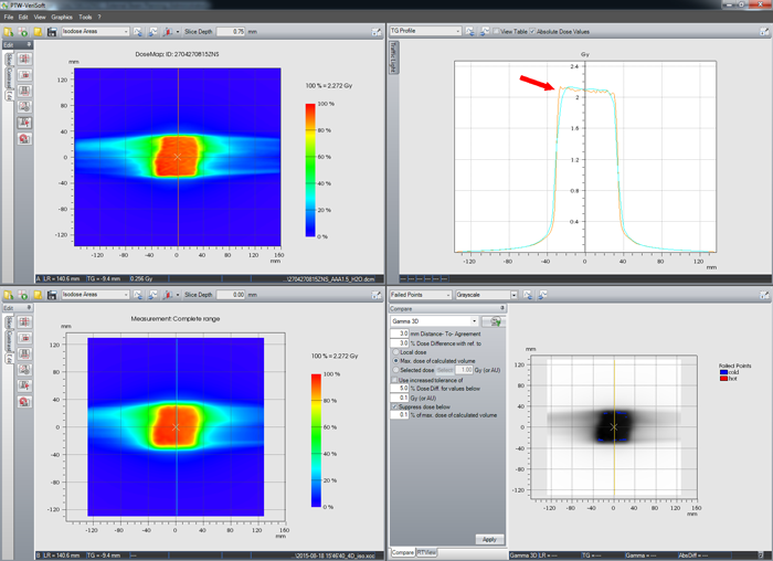
The following plan is a 6FFF glioma plan with two half-arcs. AAA calculation was done with 1.5 mm for testing purposes (the clinical plan was calculated with 2.5 mm). In the sagittal plane, the orange dose profile is much steeper than the cyan profile from measurement:
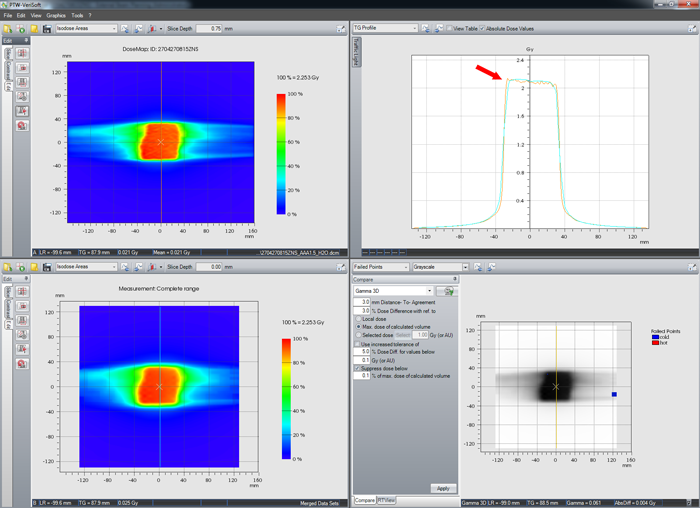
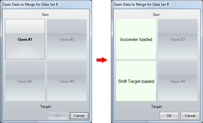
If a second measurement is performed with a shifted phantom (the shift is expected to be 5 mm away from the gantry), the measurements can be merged by selecting "File > Data Set B > Merge ...".
The higher resolution has a positive effect not only on the profiles,
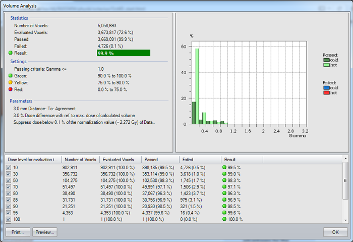
but also on the Volume Analysis results. First non-merged:
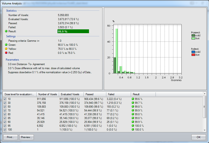
then merged:
