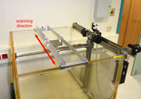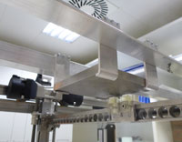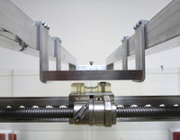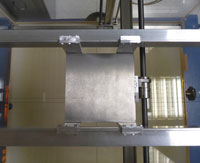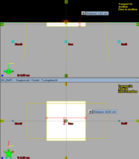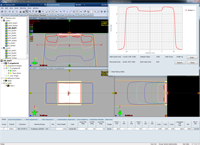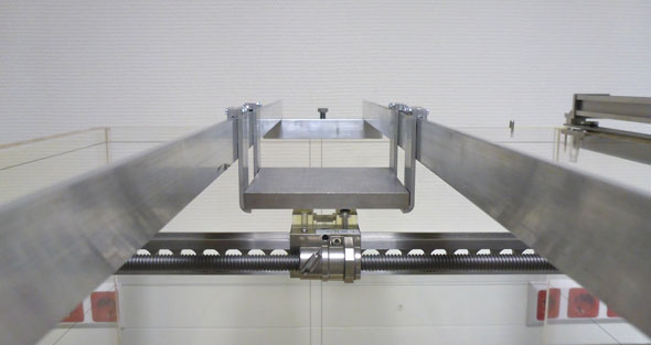
(The bridge-like construction allows continuous dose scanning in water behind a titanium absorber.)
Introduction
The accurate modelling of tissue heterogeneities during radiotherapy treatment planning is one of the key features of modern dose calculation algorithms. In the previous study on the commissioning of Acuros XB ("AXB") version 10, we used the 2D-Array for measuring 2D dose distributions behind an inhomogeneity phantom. There are several problems with this approach.
- Low spatial resolution, related to the 2D-Array.
- Inhomogeneities introduced by the measuring system itself (air-filled ionization chambers).
- Automatic conversion of the phantom's synthetic materials (PTFE, Polystyrene, PMMA, ...) to mixtures of different human tissues based on a HU to mass density calibration curve.
We think the first two points are clear, but the third point needs some explanation.
How Acuros Determines the Materials in the Patient
When Eclipse prepares for Acuros dose calculation, it first reads the HU values of the voxels1 in the CT image. This "HU image" is converted to a "mass density image" using the mass density calibration curve of the CT scanner, which has to be provided by the user. The mass density is then converted to a mixture of five types of tissue in the human body (lung, adipose tissue, muscle, cartilage, bone) plus air.
The following data is taken from the AcurosXB-11.0 Physical Materials table (there is no new AcurosXB-13.0 table in ARIA 13):
| ID | Name | Min. g/cm3 | Max. g/cm3 | Def. g/cm3 | AutoHU2Mat |
|---|---|---|---|---|---|
| Air | Air | 0.0000 | 0.0204 | 0.0012 | Yes |
| Lung | Lung | 0.0110 | 0.6242 | 0.2600 | Yes |
| Adipose | Adipose Tissue | 0.5539 | 1.0010 | 0.9200 | Yes |
| Muscle | Muscle Skeletal | 0.9693 | 1.0931 | 1.0500 | Yes |
| Cartilage | Cartilage | 1.0556 | 1.6000 | 1.1000 | Yes |
| Bone | Bone | 1.1000 | 3.0000 | 1.8500 | Yes |
These six materials are the only ones for which an automatic HU to material conversion ("AutoHU2Mat") is performed by Eclipse.
Example: if a voxel with a CT value of -483 HU is encountered, our mass density calibration curve will convert this to 0.534 g/cm^3, and this will be classified as "lung" material (although the real tissue is not always lung). A CT value of 734 HU on the other hand will be converted2 to 1.446 g/cm^3, a mixture of 31% cartilage and 69% bone etc.
Up to 3 g/cm^3 (the maximum density of "Bone"), the conversion will happen automatically. Beyond this value, manual material assignments to structures have to be performed. This was already described previously.
And in a Phantom?
If a CT-scanned phantom with inserts of various densities is used and Eclipse is allowed to determine materials automatically, dose deviations may occur. The HU value of a voxel does not say anything about the chemical composition of the material, especially if "exotic" phantom materials are used. Two materials may give the same HU numbers, although the chemical compositions (and therefore the atomic interactions) are different.
For dose calculation inside or behind a block of, say, PTFE ("Teflon"), it makes a difference whether the automatic HU to material conversion is used by the algorithm, or the material "PTFE" is assigned to the block. If the material of the block is known and one is lucky to find it in the Physical Materials Table of ARIA, it is better to assign the material to the structure, at least in the case of Acuros.
Here the data for some typical phantom materials, again taken from the AcurosXB-11.0 Physical Materials table:
| ID | Name | Min. g/cm3 | Max. g/cm3 | Def. g/cm3 | AutoHU2Mat |
|---|---|---|---|---|---|
| Polystyrene | Polystyrene | 0.5900 | 1.0750 | 1.0500 | No |
| PMMA | PMMA | - | - | 1.1900 | No |
| PTFE | PTFE | - | - | 2.2000 | No |
| Ti6Al4V_ELC | Titanium Alloy | 3.5600 | 6.2100 | 4.4200 | No |
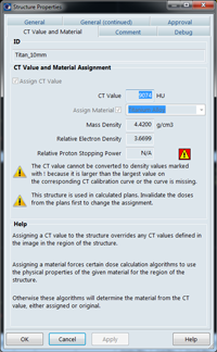
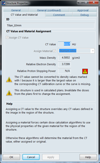
We assign the alloy Ti6Al4V_ELC (ELC stands for extra low chlorine) to our grade 2 titanium block, although this is in fact not entirely correct. The biomaterial Ti6Al4V contains 90% titanium, 6% aluminium and 4% vanadium. It contains small amounts of Fe (max. 0.25%) and O (max. 0.2%). Pure titanium on the other hand has a slightly higher density, 4.50 g/cm^3. Luckily, since the table allows for a cetrain density range between 3.56 and 6.21 g/cm^3, we can "tweak" the HU in order to get to the right density of 4.50 g/cm^3 (see sidebar).
Results
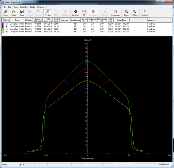
The raw dose profiles with and without titanium look like this (10FFF, two depths):
Instead of comparing measured and calculated raw dose profiles directly, we first determined the absorption profiles (difference between the attenuated and the unattenuated profile), and compared these.
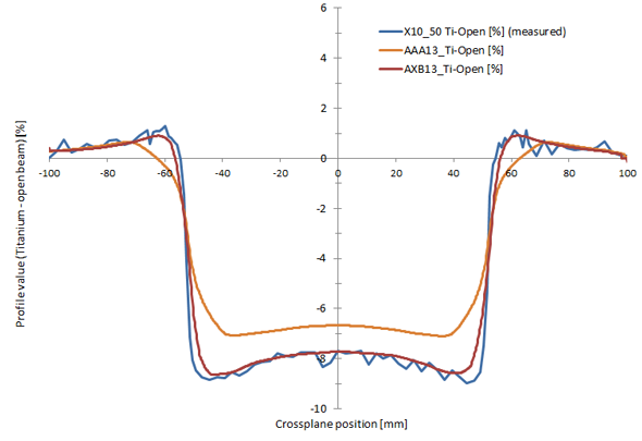
The following plot shows the measured and calculated absorption profiles behind the titanium block for 10 MV photons, SSD100, and 50 mm depth:
Absorption at central axis is about 8 percent. While AAA13 underestimates the attenuation a little, the agreement between AXB13 and measurement is excellent.
Penumbra at the edges of the block appears to be a little too broad, but the reason is probably the calculation step size of 2 mm (it was not practical to use smaller AXB calculation step size in Eclipse, due to long calculation times and high memory consumption).
The results for 15 MV and 10FFF, two more "typical" energies for pelvic region, are very similar.
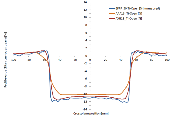
For 6FFF (the "softest" of the five photon energies), the largest attenuation values are expected. Again, AXB agrees better with measurement than AAA:
However, 6FFF will probably never be used for the pelvic region, even less if a titanium hip implant is present.
Notes
1We speak of voxels meaning a pixel with a certain "depth" (slice thickness).
2Remember that the values are only valid for our own mass density calibration curve.
