DirectDensity® and iMAR: First Results
DirectDensity® and the Iterative Metal Artifact Reduction algorithm (iMAR) are two features of our new CT. While DirectDensity (DD) aims to remove the kV-dependence of image HU which enables the clinician to improve structure delineation by choosing lower kV settings than the strict 120 kV TPS calibration voltage, iMAR reduces the intensity of metal artifacts. The implementation details of both DD and iMAR can be found in the literature1-4.
Since we are currently in the process of setting up all our scan protocols, we wanted to quickly check whether DD works properly and whether iMAR has an influence on image HU.
Materials and Methods
The CIRS 062 Electron Density Phantom (see our description from 2013) has material inserts ranging from Lung Inhale (mass density: 0.195 g/cm3) to Titanium. H2O is represented by a water filled syringe.
We scanned the phantom in the following configuration:
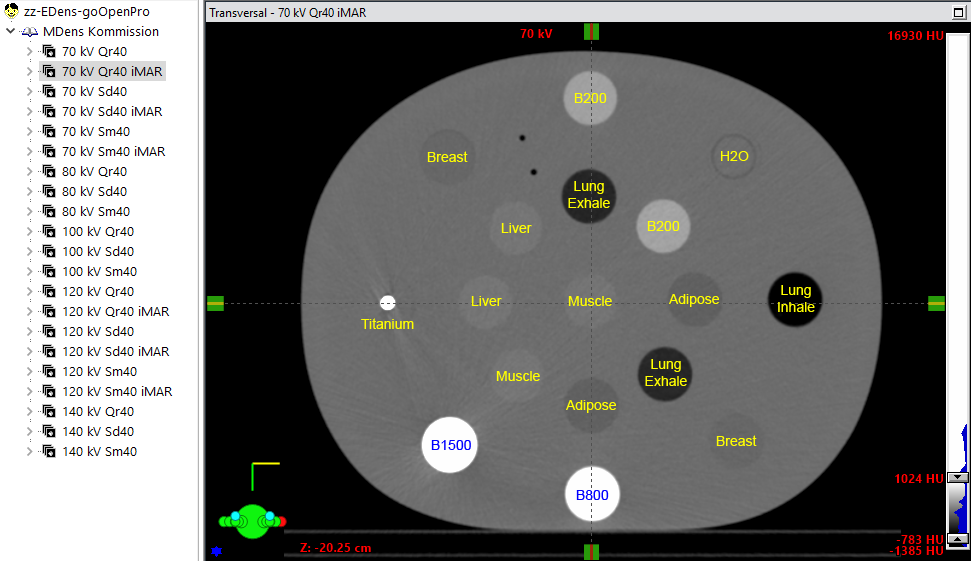
(The CIRS phantom as used in this study. The reconstructions are listed on the left.)
For scanning, we used the Direct Density Commissioning Protocol of the go.OpenPro. The protocol contains 5 spiral scans with different kV settings (70 kV, 80 kV, 100 kV, 120 kV, 140 kV). Each scan is reconstructed with three different kernels. The Qr40 is a standard RTP kernel with the typical kV dependence of image HU. The Sd40 and Sr40 kernels are DD kernels. According to Siemens, Sd40 should be selected if the TPS dose calculation algorithm (e.g., AAA) uses an electron density calibration (EDens). The Sm40 kernel on the other hand should be chosen for algorithms which use if mass density (MDens) calibration, e.g. Acuros XB or Monte-Carlo algorithms.
In order to assess the potential impact of iMAR on HU, we added additional iMAR reconstructions for 70 kV and 120 kV. In the chosen phantom configuration, the Titanium rod and also the dense bone (B1500, mass density 2.000 g/cm3) add some artefacts to our images.
The 21 reconstructions were exported to Eclipse and evaluated with a square ROI of 28 x 28 px for most phantom inserts (H2O: 22 x 22 px, Titanium: 5 x 5 px).
Results and Discussion
The following table lists the CT values for the 21 reconstructions and the CT number difference between the regular and the iMAR reconstructions (70 kV and 120 kV only).
For each material, Edens is the electron density relative to water. Mdens is the mass density in g/cm3.
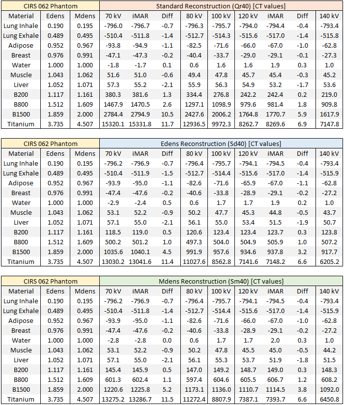
Several things can be seen from the table:
- For densities up to Liver (1.071 g/cm3), the three kernels give practically the same CT values for a selected kV setting and material, e.g. for Breast and 70 kV: -47.1 (Qr40), -47.4 (Sd40), -47.4 (SM40); for Breast and 140 kV: -27.3, -27.2, -27.2 for Qr40, Sd40 and Sm40 respectively.
- For the bone inserts (B200, B800, B1500) the situation is very different: the DD kernels Sd40 and Sm40 show very low dependence of CT numbers on kV, and the absolute numbers compared to Qr40 are different too. This is due to the DD algorithm, which provides that in this density range, the CT values ("image values") can be converted to e.g. mass density rho using the formula:

The B800 bone plug for instance has mass density 1.609 g/cm3 according to the phantom description. The CT value of the Sm40 reconstruction ranges from 601.3 (70 kV) to 606.7 (140 kV), which more or less satisfies the equation.
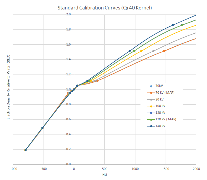
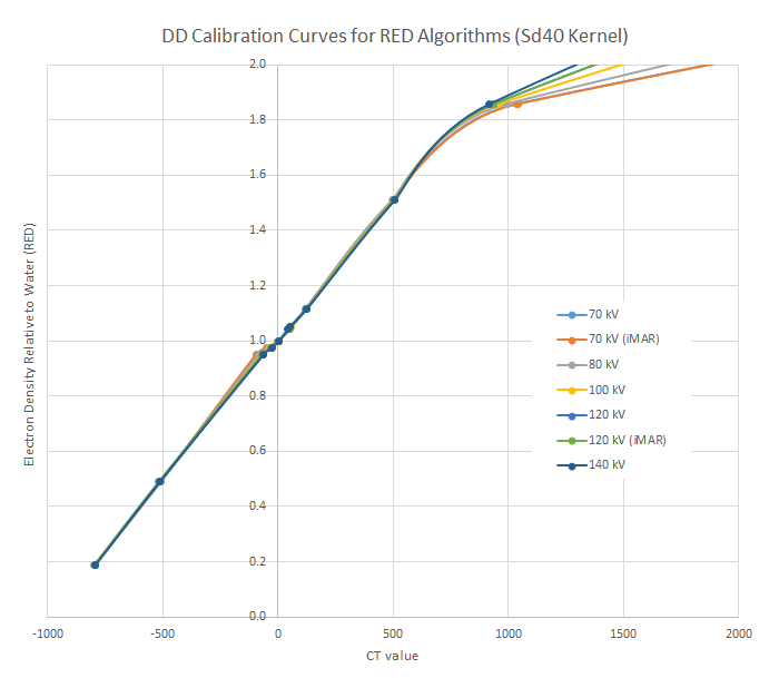
We get a better picture if we plot for the three kernels and the different kV settings the EDens or MDens curves as a function of HU (or CT number). This is the usual way the TPS calibration curves are displayed.
For the standard Qr40 kernel, bone densities typically depend on kV: the lower the voltage, the higher the HU for the same material. Note that the curves for iMAR are plotted on top of the non-iMAR curves, because they are so similar:
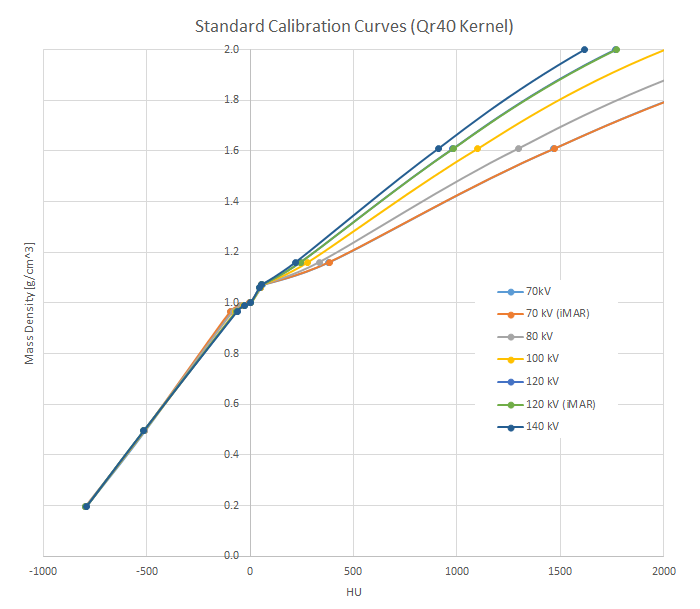
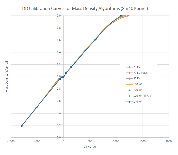
If one prefers to see the vertical axis as mass density, here is the plot.
The picture changes for the DD kernels. The effect of the DirectDensity algorithm is to bundle the curves in the bone range. Note however that beyond B1500 the curves start to diverge again, because the Titanium point is outside the scope of DirectDensity:
We plot the Sm40 kernel with mass density on the vertical axis:
 Conclusion
DirectDensity® offers the possibility of high image contrast (for contouring) and HU fidelity (for dose calculation) at the same time. However, DD scan protocols would contain two recon jobs:
- For contouring, the low kV scan (e.g., 80 kV) is reconstructed with the Qr40 kernel,
- dose is calculated on the (more or less kV-independent) DD reconstruction (Sd40 or Sm40).
DirectDensity does what it should do, in the density range it was designed for. However, when tube voltage is reduced from 140 kV to 70 kV, all three kernels roughly double the Titanium CT numbers (see table). DirectDensity is simply not made for Titanium and beyond. CT numbers >40000 are possible in patient images (gold fillings, hip implants, etc.), and the impact on dose calculation in the treatment plan is not easily assessable. It depends on the individual situation, e.g., on the volume of high density material in the beams. Although dose inside the dense inhomogeneities is rather irrelevant, the attenuation of the dense materials should be correctly taken into account. Eclipse can currently process CT numbers up to 32767 HU, which means that a Gold point can be added to calibration curves, to avoid warnings in Eclipse. If Acuros is used, a material assignment of high density voxels above 3.0 g/cm3 is mandatory anyway.
Fortunately5, starting with Eclipse 18.1, more than one calibration curve can be configured: In version 18.1, up to 20 separate electron density and mass density calibrations can be added, which means that one can have all the above kV settings calibrated separately, from Air to Gold, plus an additional DD calibration, if desired. Using a separate calibration curve for each kV setting would have two advantages:
- Only one reconstruction is needed for both contouring and dose calculation, because each kV setting has it's own calibration curve.
- The Titanium point can be correctly calibrated for each kV setting.
Concerning iMAR, we have seen that the impact on CT numbers is so low, that it does not seem to require separate TPS calibrations with and without iMAR.
Notes
1 White paper DirectDensity®, Technical principles and implications for radiotherapy, Dr. André Ritter, Dr. Nilesh Mistry, Siemens Healthineers.
2 DirectDensity cookbook, A guide to personalized CT imaging in RT, Siemens Healthineers.
3 White paper Evaluation of the Direct Density Algorithm for Energy-Independent Radiotherapy Treatment Planning, Alisha Shutler et al., Siemens Healthineers.
4 White paper Iterative Metal Artifact Reduction (iMAR), Technical principles and clinical results in radiotherapy, Marc Kachelrieß (DKFZ), Andreas Krauss, Siemens Healthineers.
5 Our ARIA and Eclipse 18.1 upgrade is scheduled for March 21, 2025.